Figure 3.
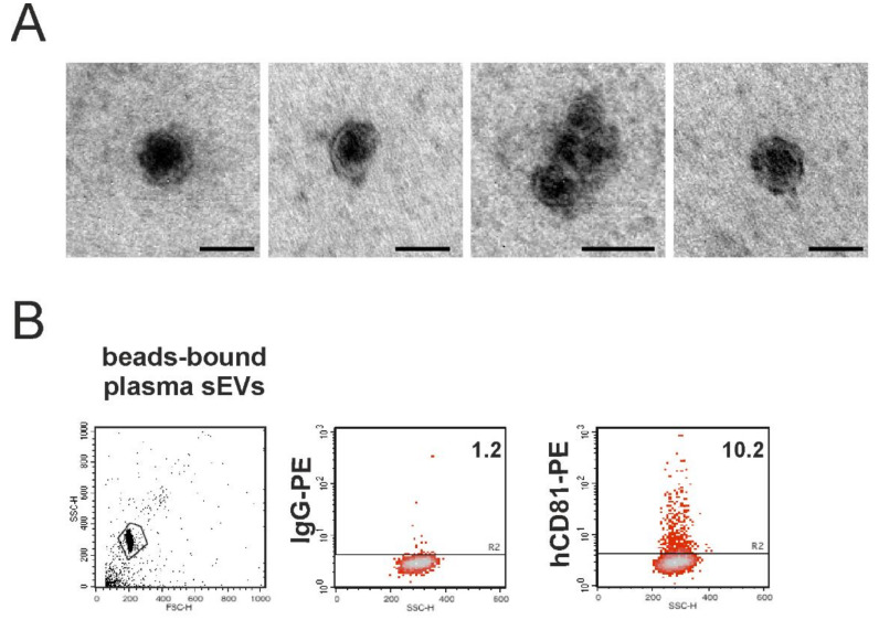
Detection of human sEVs in the mouse plasma. (A) TEM analysis of the sEVs purified from the mouse plasma. Bars, 100 nm when not indicated. (B) sEVs purified from the mouse plasma were immunocaptured by the magnetic beads conjugated to human CD63 tetraspanin and then processed for immunofluorescence of the human CD81-PE. Aggregates and debris were excluded (gating) from the fluorescence analysis, as shown in the cytogram relative to the light scatter parameters (left panel). The number reported in each cytogram represents the percentage of positivity.

