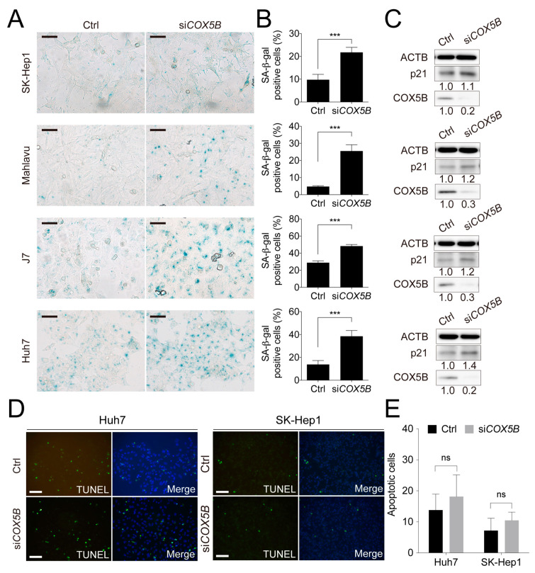
Figure 3.
Knockdown of COX5B suppresses cell proliferation through induction of cell senescence but not programmed cell death. This figure was used to supplement Figure 2 in the main text. (A) The beta-galactosidase staining assay to examine the status of cell senescence. The black Scale bar represented 50 μm. The quantitative results were shown in (B). ***, p < 0.001. (C) The western blot analysis of cell senescence marker p21 levels in HCC cells with or without depletion of COX5B. The western blot images in this panel were all acquired by using chemStudio PULS imaging system. (D) The TUNEL assay to determine the status of cell apoptosis. The white scale bar represented 50 μm. The quantitative results were shown in (E). All the in vitro cell-based assays shown in this figure were conducted in duplicates.

