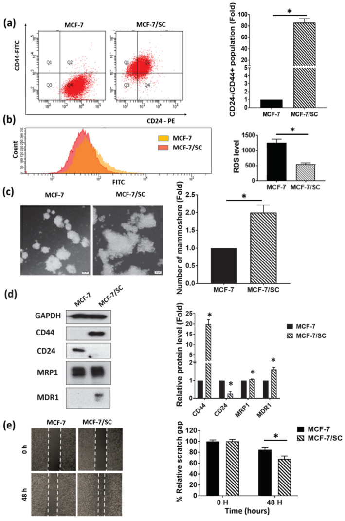
Figure 1.
MCF-7/SC exhibit more prominent cancer stem cell characteristics than the parental MCF-7 cells. (a) Fluorescence-activated cell sorting (FACS) analysis of the CD44+/CD24− cell population in MCF-7/SC and MCF-7 cells. (b) Measurement of the ROS levels in MCF-7/SC and MCF-7 cells. (c) Comparison of the mammosphere formation ability of MCF-7/SC and MCF-7 cells cultured in the MammoCult Human Medium for 10 days. Magnification 100×. (d) Analysis of the expression of cancer stem cell markers in MCF-7/SC and MCF-7 cells by Western blotting. GAPDH was used as a loading control. (e) Migratory potential of MCF-7/SC and MCF-7 cells as assessed by the wound healing assay. Data are shown as the mean ± standard deviation of three biologically independent experiments. * p < 0.05 vs. control.

