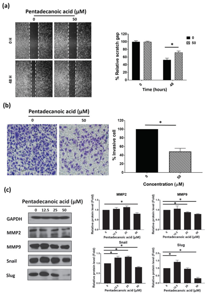
Figure 3.
Pentadecanoic acid inhibits the migration and invasion of MCF-7/SC. (a) Cell migration was determined by the wound healing assay following 48 h of exposure. (b) Invasive cells were stained with crystal violet after treatment with pentadecanoic acid for 48 h (magnification 100×). (c) Western blot analysis of epithelial–mesenchymal transition (EMT) markers in MCF-7/SC was performed after pentadecanoic acid treatment for 48 h. GAPDH was used as a loading control. Data are shown as the mean ± standard deviation of three biologically independent experiments. * p < 0.05 vs. control.

