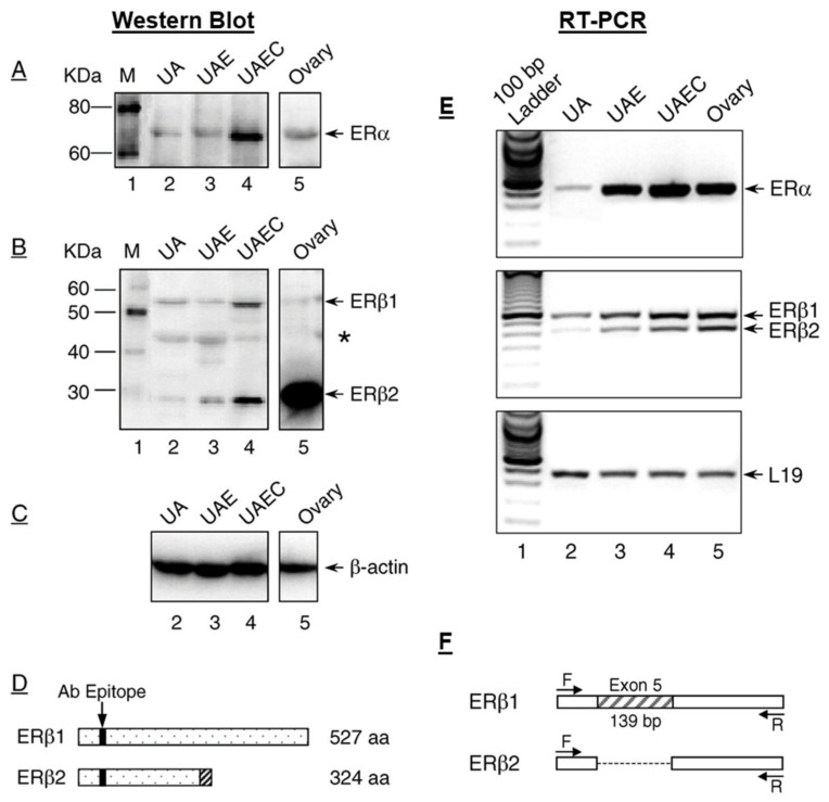
Figure 1.
Uterine artery estrogen receptors. Estrogen receptor (ER)α (A) and ERβ (B) proteins in the protein extracts of intact uterine artery (UA), purified UA endothelium (UAE), cultured UA endothelial cells (UAEC), and ovary from pregnant ewes detected by Immunoblotting with epitope specific antibodies. β-actin (C) was used as loading control. (D) A diagram representing the truncated form of ERβ2 that results from the splicing deletion of exon 5. The shadowed box represents the amino acid sequences encoded by different reading frame. (E) ERα and ERβ mRNAs detected by RT-PCR with the ribosomal protein L19 as loading control. (F) A diagram shows a 139bp deletion of Exon 5 in ERβ2 mRNA vs. the native ERβ1. Uterine artery (UA); UA endothelium (UAE); UA endothelial cells (UAEC); amino acid (aa). Bands marked with * may indicate additional truncated forms of ERβ. Adopted from Liao et al. [71].

