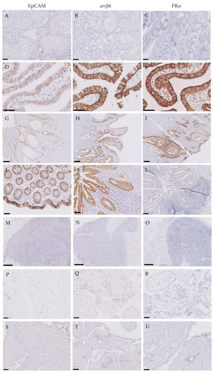
Figure 2.
Expression patterns of epithelial cell adhesion molecule (EpCAM), integrin αvβ6 and folate receptor-α (FRα) in tumor-negative tissues. Representative images of immunohistochemically stained tissue samples of tumor-negative ovaries (A–C), fallopian tubes (D–F), endometrium (G–I), intestine (crypt) (J–L), lymph nodes (M–O), omentum (P–R) and peritoneum (S–U) for EpCAM, αvβ6 and FRα. Scale bars represent 50 µm (A–C, K–L), 100 µm (J, M–U), 200 µm (D–F) and 500 µm (G–I).

