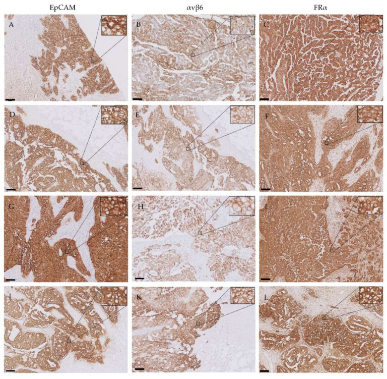
Figure 4.
Representative images of primary tumors and their corresponding omental, peritoneal and lymph node metastases immunohistochemically stained for EpCAM, αvβ6 and FRα. The following structures are shown: primary ovarian tumors (A–C), omental metastases (D–F), peritoneal metastases (G–I) and lymph node metastases (J–L). All images include tumor tissue showing positive expression and adjacent healthy tissue showing no expression. While EpCAM and FRα display high intensity staining in all tumor cells, αvβ6 exhibited more heterogeneous staining intensities in these cells. Scale bars represent 100 µm. Inserts show tumor cells at a higher magnification.

