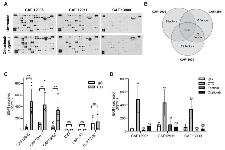
Figure 3.
Cetuximab treatment alters CAF secretion profiles. (A) Raw images of cytokine array blots performed on conditioned media collected from CAFs treated with IgG control or 1 μg/mL cetuximab for 72 h. Boxed readings indicate epidermal growth factor (EGF). (B) Cytokine and growth factor expression was evaluated via cytokine arrays. The overlap of upregulated cytokines (>0.5 fold compared to untreated) across CAF lines is shown. (C) Levels of EGF secretion were determined via enzyme-linked immunosorbent assay (ELISA) on primary CAFs (12905, 12911, 13000), cancer cells (DiFi, LIM1215), and normal primary fibroblasts (NCF12737) lines. (D) Conditioned media was collected from CAFs treated with 1 μg/mL cetuximab, 1 μM erlotinib, 5 μM oxaliplatin, or 1 μg/mL IgG control for 72 h. ELISAs were performed to evaluate levels of EGF. p-value ≤ 0.01: **; p-value ≤ 0.05: *; ns: not significant.

