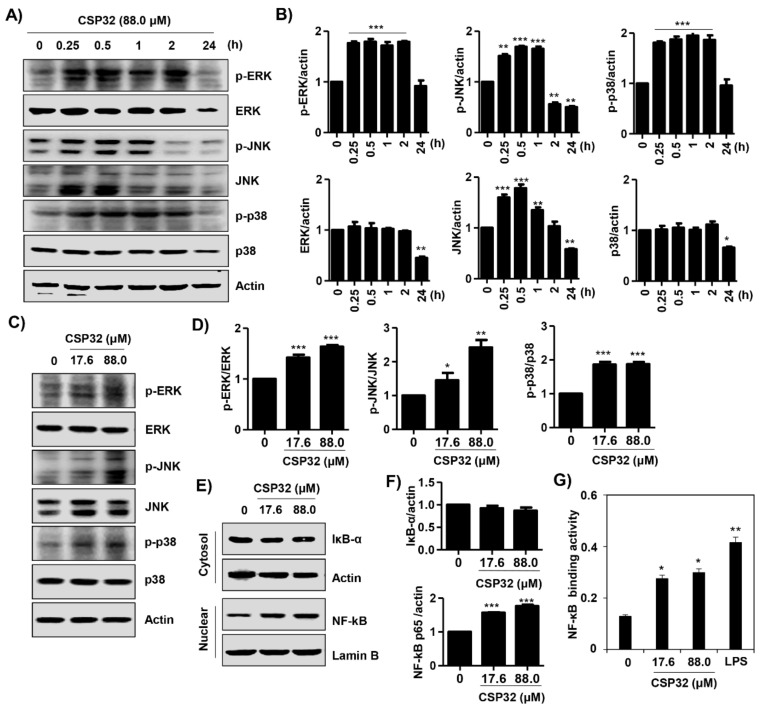
Figure 4.
CSP32 activated MAPKs and the NF-κB signaling pathway in macrophages. (A) At 0, 15 min, 30 min, 1 h, 2 h, and 24 h after CSP32 treatment, cells were harvested and lysed. (C,E) Cells were treated with the indicated concentration of CSP32 for 1 h and subsequently harvested and lysed. (A,C) Total cell lysates were examined by western blotting for ERK, JNK, and p38 MAPK phosphorylation. β-actin was used as an internal control. (E) Cytoplasmic and nuclear lysates were examined by western blotting for IκB-α and NF-κB. Actin and lamin B1 serve as the internal controls for the cytoplasmic and nuclear lysates, respectively. (B,D,F) Quantitative analysis of protein expression. The expression of each protein was indicated as a fold change relative to the control. Data are expressed as the mean ± SD (n = 3). * p < 0.05, ** p < 0.01 and *** p < 0.001 compared with the control. (G) In nuclear lysates, NF-κB activity was analyzed using an NF-κB p65 transcription factor assay kit. Data are expressed as the mean ± SD (n = 4). * p < 0.05 and ** p < 0.01 compared with the control.

