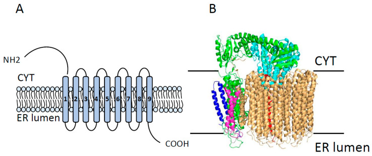
Figure 4.
Overall structure of the V0 domain of V-ATPase. (A) Topology of V0 domain showing its 9 transmembrane segments with large cytoplasmic N-terminal and C-terminal domains. (B) The overall architecture of the V0 domain of V-ATPase (Protein Data Bank [PDB] code 6C6L), showing all known components of the V0 domain, including subunits a (in red), d (in cyan), e (in blue), f (in pink), and the c-ring (in wheat). CYT = cytoplasm; ER lumen = endoplasmic reticulum lumen. Modified from Roh et al. 2018.

