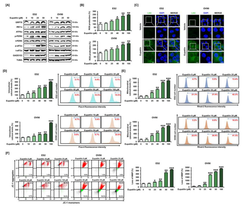
Figure 2.
Effects of eupatilin on various aspects of cellular stress in ovarian cancer. (A) Western blot of endoplasmic reticulum (ER) stress regulatory proteins after ES2 and OV90 cells were treated with different concentrations of eupatilin. (B) The effects of eupatilin on reactive oxygen species (ROS) generation in ES2 and OV90 cells were evaluated by flow cytometry with dichlorofluorescin (DCF) fluorescence signals. (C) The effect of eupatilin on lipid peroxidation was determined by immunocytochemistry of linoleamide alkyne (LAA) to indicate lipid peroxidation with green fluorescence in the cytosolic fraction in ES2 and OV90 cells. The scale bar indicates 20 μm. (D–E) Eupatilin-mediated intracellular (D) and mitochondrial (E) calcium levels were investigated by flow cytometry with Fluo-4 and Rhod-2 fluorescence signals, respectively, after eupatilin treatment in ES2 and OV90 cells. (F) The mitochondrial membrane potential (MMP, ΔΨm) was analyzed by the distribution of red and green fluorescence using JC-1 staining after eupatilin treatment in ES2 and OV90 cells. The experiments were performed in triplicate. Data represent the mean ± standard deviation, and asterisks indicate that the effect of treatment was statistically significant (* p < 0.05, ** p < 0.01, and *** p < 0.001). Detailed information about the western blot can be found in Figure S1.

