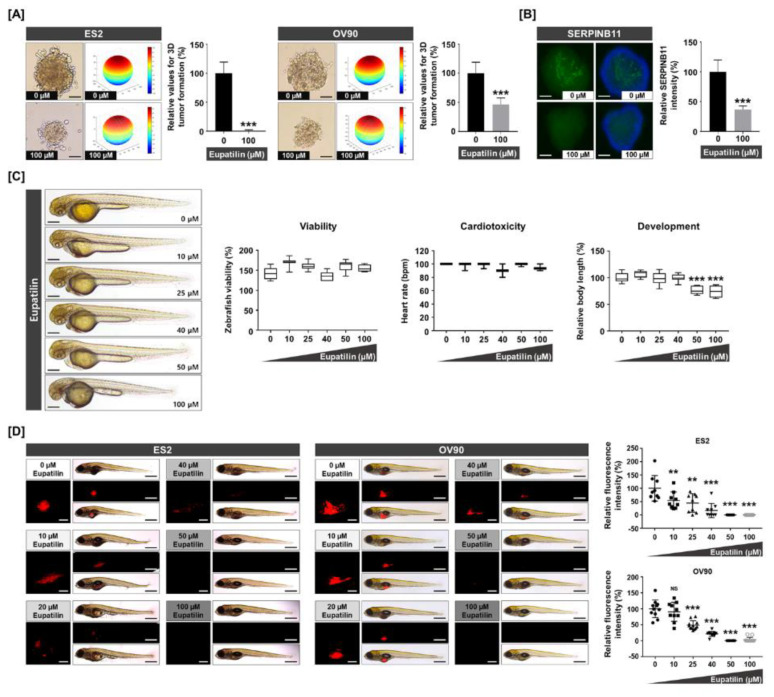
Figure 7.
Anticancer effects of eupatilin on in vitro and in vivo 3D anchorage-independent growth and xenograft models. (A) The 3D tumor spheroid-forming capacity shows that eupatilin exhibits anticancer potential under anchorage-independent cell growth conditions. The scale bar indicates 20 μm. (B) Immunofluorescence of SERPINB11-positive cells (green), which were counterstained with DAPI (blue) on the ES2 tumor spheroid. The scale bar indicates 20 μm. (C) Zebrafish embryos of 24 hpf were treated with increasing concentrations of eupatilin for 24 h to analyze morphology, viability, cardiotoxicity, and development as toxicological parameters. The scale bar indicates 250 μm. (D) In vivo assay of anticancer effects by eupatilin in a zebrafish xenograft model. Red fluorescence intensities were quantified to determine tumor formation. The scale bar indicates 25 μm in square panels and 100 μm in rectangle panels. The experiments were performed in triplicate. Data represent the mean ± standard deviation, and asterisks indicate that the effect of treatment was statistically significant (** p < 0.01 and *** p < 0.001).

