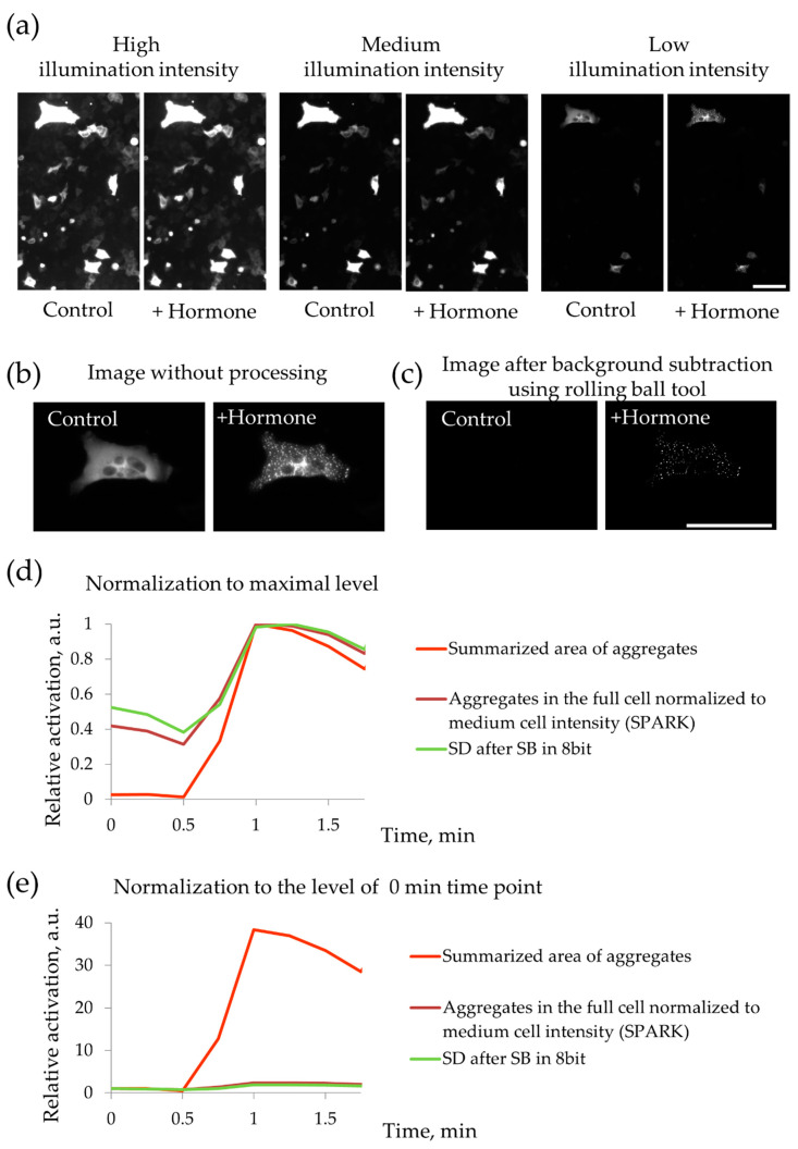
Figure 5.
Testing the method of registration of PKA activity using PKA-Spark fluorescent probe. (a) PKA-Spark was expressed in HeLa-Kyoto cells with different expression levels and the cells were stimulated with serotonin (10−5 M). Fluorescence of the same field of view was registered with different illumination intensity (marked above the pictures). It is seen that only the most bright cells expressing the probe with relatively high level, demonstrated hormone-dependent response. Scale bar 100 µm. (b,c). Processing of fluorescent images of responding cells. (b) Responding cell before and after hormone adding without image processing. (c) Fluorescent figures after background subtraction using rolling-ball tool. Scale bar 100 µm. (d,e) Test of different strategies of quantification PKA-Spark fluorescent droplets formation. (d) The results of quantitative analysis were normalized to the maximum measured level. (e) The results of quantitative analysis were normalized to the zero time point.

