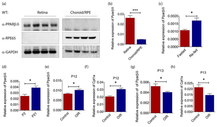
Figure 1.
Peroxisome proliferator-activated receptor (PPAR)β/δ is highly expressed in mouse retina. (a) Representative Western blot of PPARβ/δ, retinal pigment epithelium-specific protein 65 kDa (RPE65) and glyceraldehyde-3-phosphate dehydrogenase (GADPH) in the retina (n = 4) and choroid/RPE (n = 4) compartments of adult C57BL/6 mice. Relative gene expression of Pparβ/δ in (b) the retina (n = 10) and choroid/RPE (n = 3) compartments of C57BL/6 mice; (c) the retina of fasted (n = 4) and re-fed (n = 4) adult C57BL/6 mice; (d) the retina of P2 (n = 4) and P21 (n = 4) C57BL/6 mice, as determined by quantitative real-time polymerase chain reaction (RT-qPCR) analysis. Relative expressions of (e) Pparβ/δ and (f) Carnitine palmitoyltransferase 1A (Cpt1a) in P12 hyperoxic retina of C57BL/6 mice subjected to oxygen-induced retinopathy (OIR) (n = 4) as compared to those in age-matched normoxic retina (n = 4), as determined by RT-qPCR analysis. Relative expressions of (g) Pparβ/δ and (h) Cpt1a in P13 hypoxic retina of C57BL/6 mice subjected to OIR (n = 4) as compared to those in age-matched normoxic retina (n = 4), as determined by RT-qPCR analysis. Gene expressions are quantified relative to the housekeeping gene, Gadph, as detailed in the Materials and Methods. Data are expressed as mean ± standard error of the mean (SEM). Unpaired, two-tailed t-test was used for statistical analysis; *** p < 0.001, * p < 0.05.

