FIGURE 1:
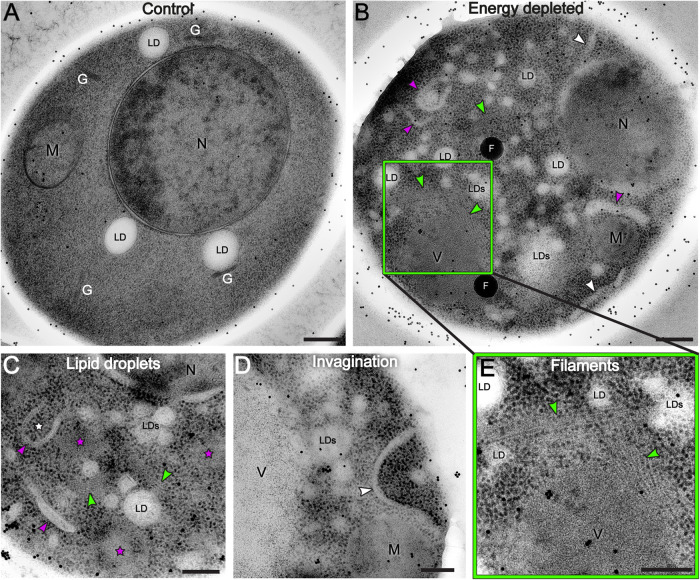
TEM images reveal cytoplasm reorganization in dormant yeast cells. A log-phase growing yeast cell (A) is compared with energy-depleted yeast cells (B–D) showing that the cytoplasm undergoes drastic reorganization upon sudden energy depletion. The vacuole is not visible in panel (A) because it is not included in this particular section of the cell. The dispersed black dots in the images are gold beads of 15 nm diameter used as alignment fiducials during tomography reconstruction. (B–D) Energy-depleted cells contain numerous fragmented lipid droplets (LDs; smaller droplets are not labeled), nascent autophagosomes (white stars), and elongated membranous structures (magenta arrows). Ribosomes appear more densely packed in energy-depleted cells than in the control cell, and areas of ribosome exclusion are also visible (magenta stars in C). (B,C) Elongated invagination of the cell membrane (white arrows) (B,D) and filamentous structures are visible in several parts of the cell (green arrows) (B,C, inset E). N = nucleus; M = mitochondria; G = Golgi; LD(s) = lipid droplet(s); V = vacuole; F = fiducial beads. Scale bars: A and B = 300 nm; C–E = 200 nm.

