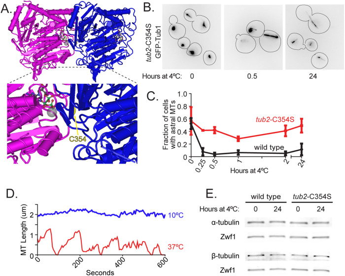
FIGURE 5:
Preventing tubulin maturation stabilizes microtubules at low temperatures. (A) Model of tubulin heterodimer with arrow pointing to residue C354 on β-tubulin (PDB entry 5W3H; Howes et al., 2017). (B) Representative field of tub2-C354S cells expressing GFP-Tub1 shifted from 30°C to 4°C for 0, 0.5, and 24 h. These images use an inverted lookup table to enhance contrast for the GFP-Tub1 signal. (C) Proportion of wild-type (black) and tub2-C354S (red) cells with astral microtubules present after shifting to 4°C for indicated time. Values are mean ± SEM from at least three separate experiments with at least 420 cells analyzed for each timepoint. (D) Lifeplots of a single tub2-C354S astral microtubule at 10°C (blue) and 37°C (red). Microtubule lengths were measured at 5-s intervals. (E) Western blot of protein lysate from wild-type (left) and tub2-C354S (right) cells incubated at 30°C and at 4°C for 24 h and probed for α-tubulin, β-tubulin, and Zwf1.

