Figure 6.
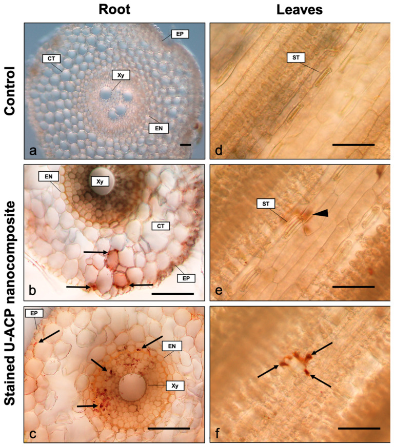
Histological localization of U-ACP nanocomposites inside wheat plants. Hand sections of wheat root (a,b,c) and leaf samples (d,e,f). Non-exposed controls show the absence of nanoparticles either in roots (a) and leaves (d). Accumulation of Alizarin Red S stained nanocomposites (arrows) can be observed in the apoplast of the root cortex (b) and inside the symplast of the central cylinder (c). In leaves, the presence of nanoparticles under the epidermis is first seen as an out of focus red-brownish stain (arrowhead) under the stomata (e), appearing as micro-sized aggregates (arrows) in the stomatal cavity when the plane is in focus (f). Note that (e) and (f) correspond to the same observation point with different focal planes. Scale bar = 50 µm. CT—cortex; Xy—xylem; EN—endodermis; EP—epidermis; ST—stomata.

