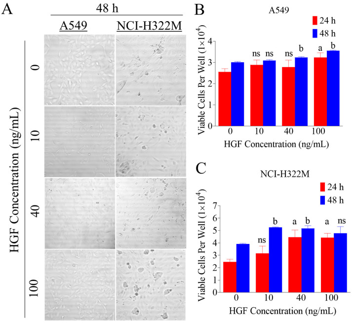
Figure 1.
Effect of increasing hepatocyte growth factor (HGF) treatments on lung cancer (LC) cells proliferation. (A) Representative image of A549 and NCI-H322M LC cells viability assay in 96 well plate with increasing HGF concentrations after 48 h. (B,C) HGF stimulated the human LC cells (A549 and NCI-H322M) proliferation in a dose-dependent manner, reaching a maximum effect at 40 ng/mL over 48 h culture period. Cells were plated at a density of 1 × 10⁴ cells/well in 96-well plates and maintained in 10% FBS supplemented media and allowed to adhere overnight. The next day, cells were washed with PBS, divided into different HGF treatment groups. Viable cells count was determined by MTT assay after 48 h. Vertical bars indicate the mean cell count ±SD in each treatment group. a p < 0.05 significantly different compared to vehicle-treated controls at 24 h and b p < 0.05 significantly different, compared to vehicle-treated controls at 48 h, ns: statically not significant.

