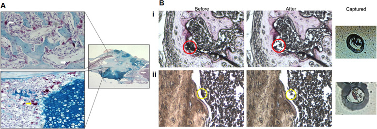
Fig. 1.
Localization of TRAP-positive chondroclasts and osteoclasts and LCM capture. a A mouse femur 12 days post-fracture stained with tartrate-resistant acid phosphatase (TRAP) (red) and counterstained with Alcian Blue, a specific stain for cartilage, shows TRAP-positive cells present in both the mineralizing cartilage (yellow arrow) and the mineralizing bone (white arrow) of the fracture callus. The TRAP-positive cells adjacent to the mineralizing cartilage are chondroclasts while those adjacent to the mineralizing bone are osteoclasts. b Seven-micrometer-thick sections cut in the frontal plane from demineralized femurs harvested 12 days post-fracture were stained for TRAP-positive cells and serially dehydrated in ascending grades of ethanol and finally xylene prior to laser capture microdissection. (i) TRAP-positive cells adjacent to mineralized cartilage (chondroclasts—red circles) are shown before LCM and after LCM, and the captured cells are shown attached to the LCM membrane. (ii) TRAP-positive cells adjacent to mineralized bone (osteoclasts—yellow circles) are shown before and after LCM, and the captured cells are shown attached to the LCM membrane

