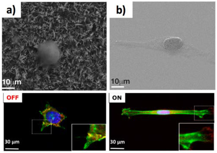
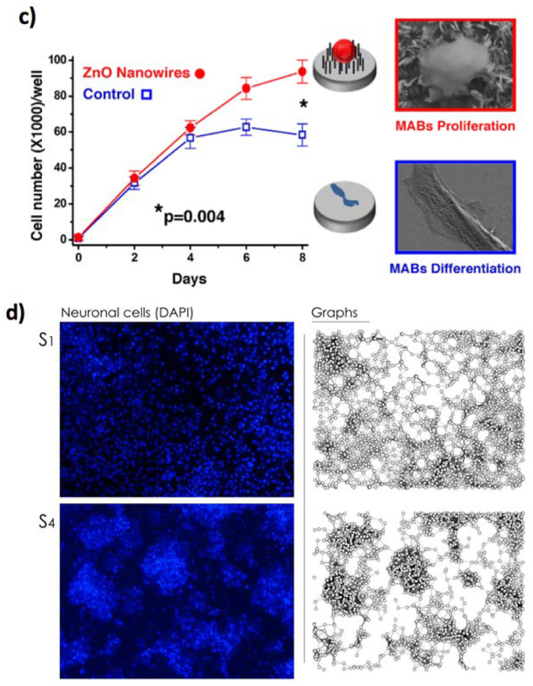
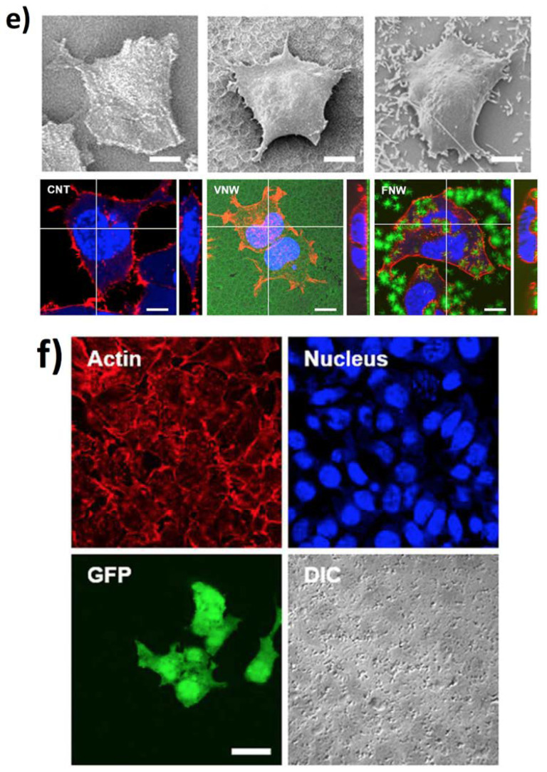
Figure 7.
ZnO NWs for regenerative medicine and drug delivery. SEM and confocal microscopy images of the MABs (a) reduced spreading onto ZnO NWs, in comparison to (b) the control glass surface. Actin is stained in green and paxillin in red. (c) The MABs proliferation on glass (blue squares) or ZnO NWs (red circles). Reprinted with permission from ref. [132]. Copyright (2018) American Chemical Society. (d) The triggering of networks formed by hippocampal neurons onto ZnO NWs. On the left, the cells imaged via fluorescence. On the right, the wiring diagrams by a Waxman algorithm. S1 and S4 indicate two different samples. Reproduced from ref. [141], under the terms of the Creative Commons CC BY license. (e) Characterization via SEM images of the HEK293 cells cultivated on glass, VNW, and FNW. Cells cultivated on NW arrays for 48 h stained for cellular nuclei (blue) and cytoskeleton (red). (f) DNA-coated on FNW is intracellularly delivered to HEK293 cells leading to a GFP-expression construct. Reproduced from ref. [144] with permission from The Royal Society of Chemistry.


