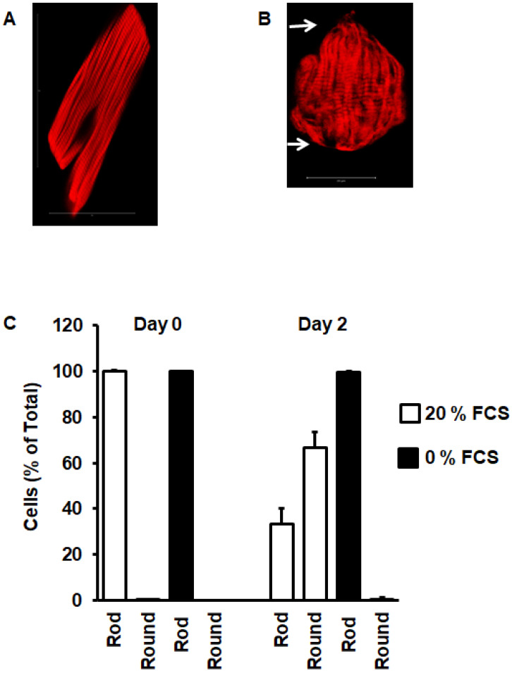
Figure 2.
Cell rounding during cultivation. (A) Freshly isolated cardiomyocyte with phalloidin staining of actin to visualize striation of sarcomeres. (B) Cardiomyocytes after 48 h cultivation in the presence of fetal calf serum, indicating sarcomere degradation from the cell poles. (C) Quantification of the number of rod-shaped cardiomyocytes and round cardiomyocytes at start of cultivation (day 0) and at day 2. Data show the effect of serum on cell rounding. Data are means ± SD from n = 8 preparations (237–410 cells per preparation were analyzed).

