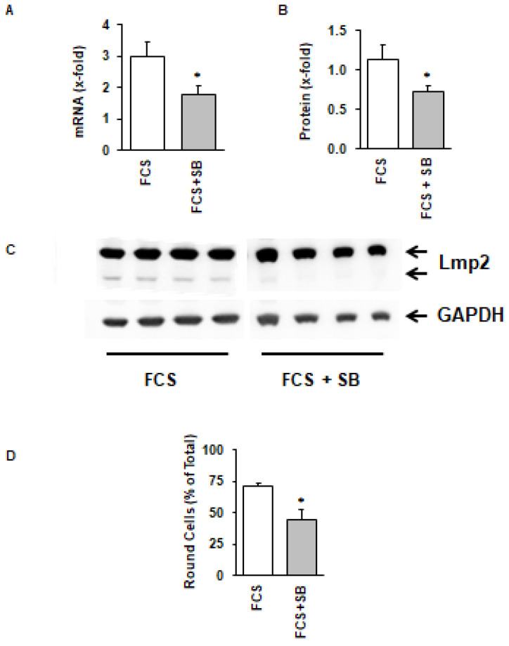
Figure 3.
Cell size (A–C) of freshly isolated cardiomyocytes with one (n = 1) or two (n = 2) nuclei (A, µm length; B, µm width; C, µm3 volume). Percent of cells to round down in the presence of fetal calf serum is shown in (D). Data are means ± SD from n = 11 preparations (96–304 cells per preparation). (*, p < 0.05 vs. FCS)

