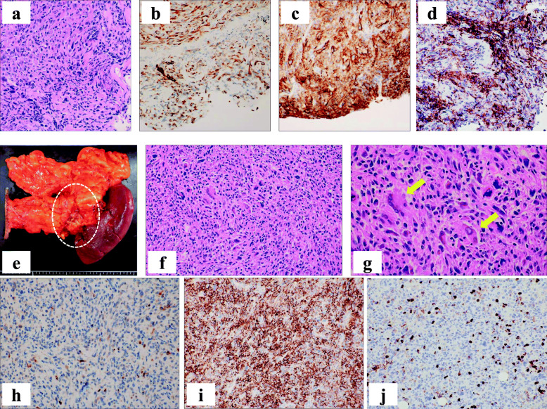
Fig. 3.
Pathological examination of the lung and pancreas. a-d Transbronchial lung biopsies: The cancerous cells show pleomorphic atypical cells containing hyperchromatic nuclei in the pulmonary lesion, forming abortive glands and solid nests with fibrous stroma (a, HE, original magnification × 200). Immunohistochemical stains show cytokeratin (CK)-7-positive (b, original magnification × 200) and programmed death ligand-1 (PD-L1) cancer cells (c, original magnification × 200). Leukocyte common antigen (LCA) is also positive in stromal cells (d, original magnification × 200). e-j Pancreatic lesion: The hard tumor is located on the ventral side of the tail of the pancreas with anterior invasion to the stomach (dotted circle in e). Sarcomatoid appearance with spindle-shaped cells and pleomorphic multinucleated cells (f, HE, original magnification × 200). Osteoclast-like giant cells (OGCs) are also seen (arrows) in the tumor (g, HE, original magnification × 400). Immunohistochemistry: CK-7 (h) and PD-L1 (i) are also positive. Small amount of lymphocytic infiltration around the cancer cells was observed in LCA staining (j, original magnification × 200)

