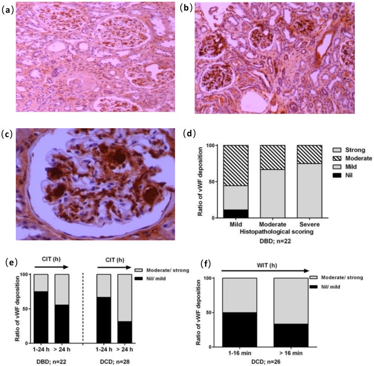
Figure 2.
Immunostaining of von Willebrand factor (vWF) and reactivity analysis in glomerular areas. Examples of subjective moderate and strong staining in reaction intensity ((a,b), 100×) and strong vWF reactivity appeared within capillary convolutes ((c), 400×). (d) vWF reactivity plotted against histological changes in DBD and DCD kidneys. (e) The effect of cold ischemic time (CIT) within 24 h or more than 24 h on vWF deposition in the DBD and DCD kidneys. (f) Semi quantitative evaluation of the DCD kidneys for vWF staining plotted against WIT.

