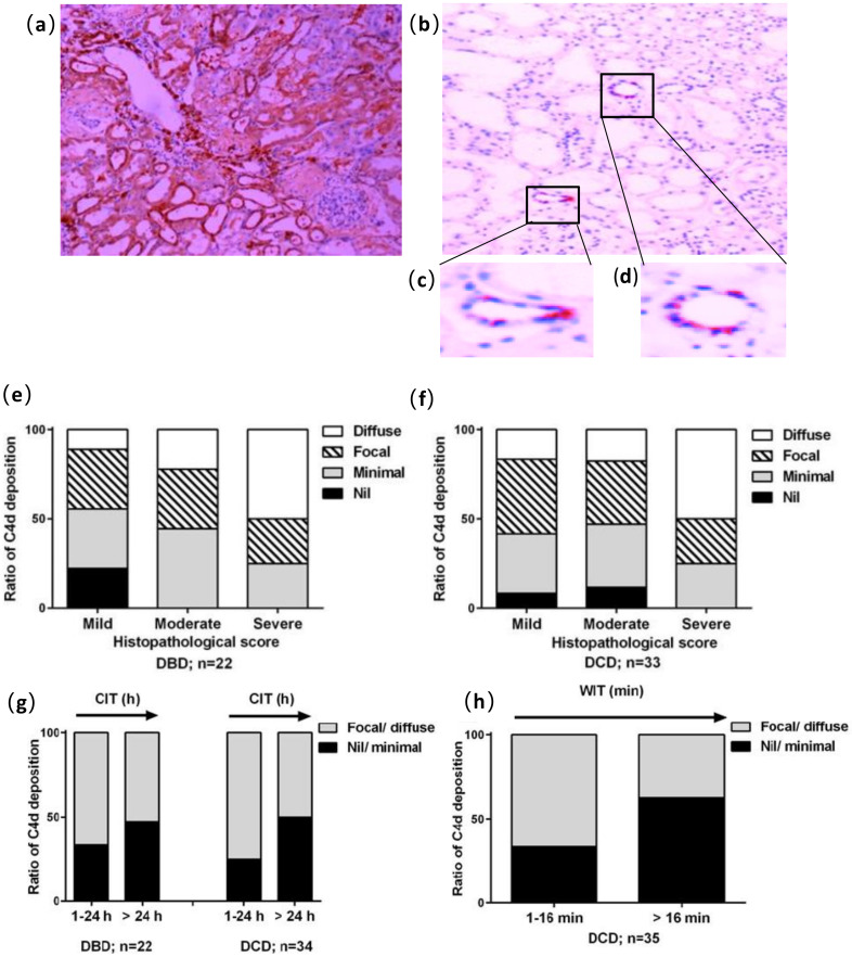
Figure 3.
Immunostaining analysis of C4d deposition in tubular epithelia (100×). (a) Diffuse deposition; (b) focal deposition; (c,d) enlarged pictures demonstrating the deposition of C4d around tubular epithelial cells; (e,f) C4d reactivity plotted against histological changes in DBD and DCD kidneys (n = 23 and 27, respectively); (g) The effect of CIT ≤ 24 h or > 24 h on C4d deposition in DBD and DCD kidneys; (h) WIT-affected C4d deposition in DCD kidneys.

