Figure 1.

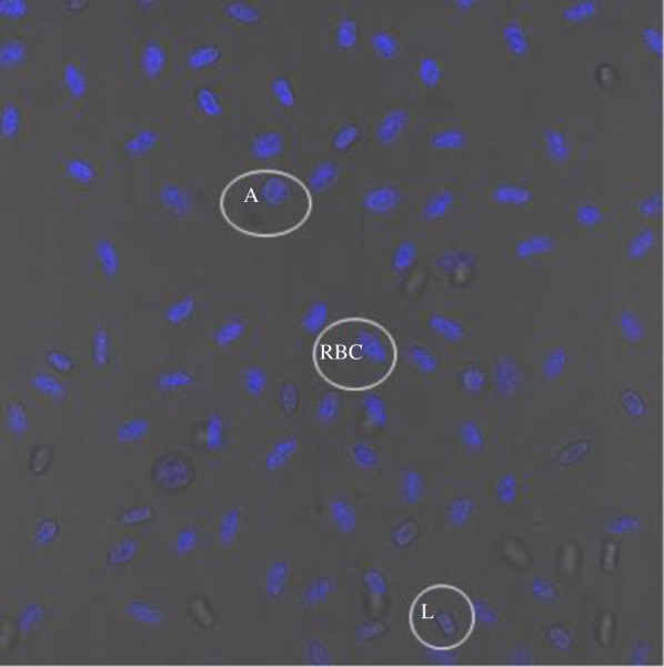
A representative confocal microscopy image of DAPI stained blood cells of Ctenophorus pictus, with azurophil (A), lymphocyte (L), and red blood cell (RBC) highlighted.

A representative confocal microscopy image of DAPI stained blood cells of Ctenophorus pictus, with azurophil (A), lymphocyte (L), and red blood cell (RBC) highlighted.