Figure 1.
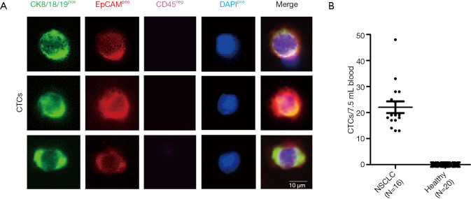
CTC analysis. (A) At the time of staging, CTCs were identified in 7.5 mL of blood by immunofluorescent staining. Presented are staining patterns of three CTCs from different NSCLC patients. (B) CTC counts in NSCLC patients and healthy never-smoking controls (bars: mean ± standard error of the mean). CTC, circulating tumor cell; NSCLC, non-small cell lung cancer.

