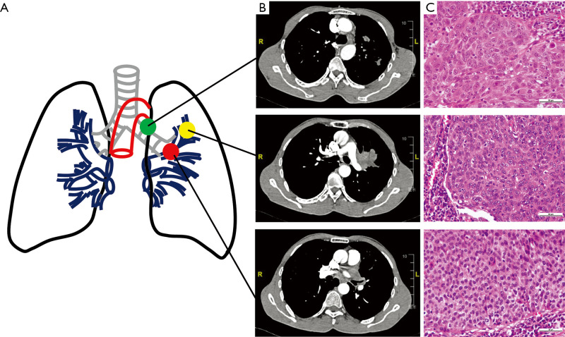
Figure 1.
Clinical and histological diagnostic results of a patient with LUSC. (A) Schematic diagram of the primary tumors (PT) and lymph node metastases (LNM) and tumor thrombus in pulmonary vein (TPV). (B) Preoperative enhanced computerized tomography (enhanced-CT) scanning showed the PT (upper), LNM (middle) and TPV (lower). (C) Postoperative paraffin section and hematoxylin and eosin (H&E) staining image based on 400× magnification. Tumor cells in PT, LNM and TPV were moderately or poorly differentiated. PT, primary tumor; LNM, lymph node metastases; TPV, tumor thrombus in pulmonary vein.

