
Figure 4. Genetic deletion or pharmacological inhibition of CypD results in stronger repaired bone in male mice at post fracture day 35.
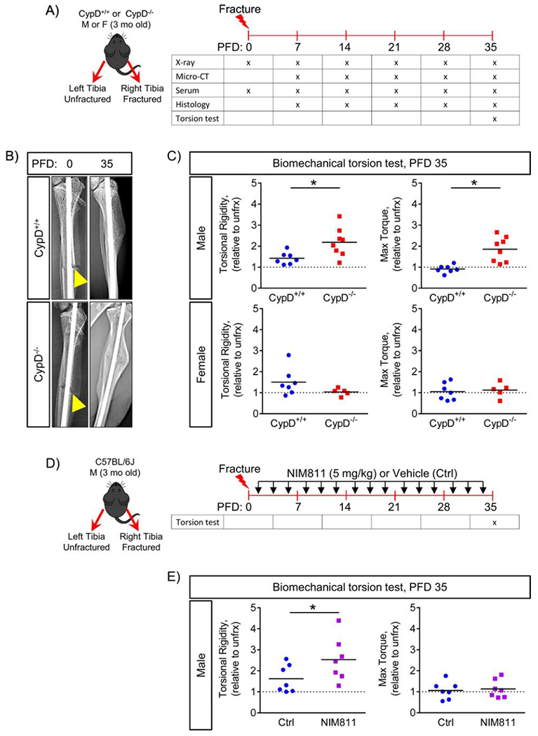
A) Tibial fractures were performed on 3-month-old male and female mice; and fractured and contralateral unfractured bones were collected for various analyses; B) Representative X-rays at post fracture day (PFD) 0 and 35. Arrowheads indicate the fracture site; C) Biomechanical properties of repaired bone were measured at PFD 35 by torsion test and reported as torsional rigidity indicating bone toughness and maximum torque indicating bone strength. Data are expressed as relative to the contralateral unfractured limb to account for differences in bone phenotype between animals. CypD−/− male mice show increased torsional properties of repaired bone; whereas, CypD−/− female mice show no difference when compared to control CypD+/+ littermates; D) Tibial fracture was induced similarly to the CypD KO mice and underwent biomechanical torsion test at PFD 35. NIM811 at 5 mg/kg or vehicle was injected intraperitoneally 3 times per week throughout the healing process; E) NIM811-treated mice show an increase in torsional rigidity and no difference in maximum torque when compared to vehicle-treated controls. Plots show actual data points and calculated means. *, p < 0.05 vs CypD+/+ controls as determined by an unpaired t-test. See also Supplementary Figure S3 & S4.