
Figure 6. Accelerated callus ossification in CypD knock-out mice.
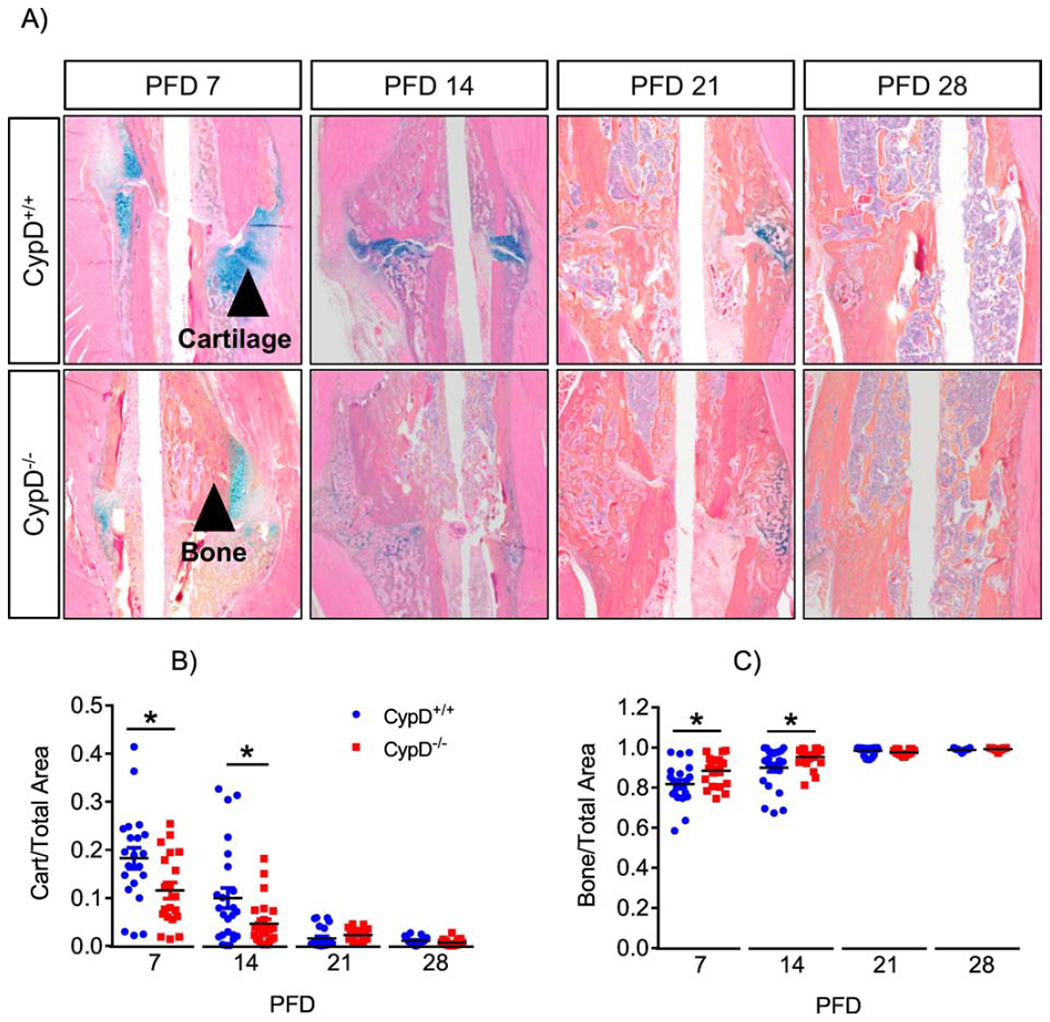
Histological staining using ABH/OG stain followed by histomorphometry of fractured bones was performed at various time points (A). CypD−/− mice show accelerated callus ossification and a truncated cartilaginous phase at PFD 7 and 14 (B & C). Plots show actual data points and calculated means. *, p < 0.05, determined by unpaired t-test of each time point, t-test was used because samples at each time point were collected and analyzed independently of other time points. See also supplementary Figure S6.