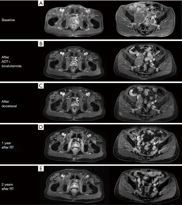
Figure 3.
Shrinkage of primary tumor and metastatic lymph nodes after the sequential therapy. Contrast enhanced axial T1-weighted MR image of the pelvis showed (A) the baseline situation of the prostate mass (44 mm × 30 mm) and bulky metastatic lymph nodes (53 mm × 77 mm, 19 mm × 29 mm); (B) obvious prostate mass regression (17 mm × 14 mm) and slight metastatic lymph node reduction (56 mm × 34 mm, 16 mm × 13 mm) after 9-month ADT treatment; (C) continual metastatic lymph nodes reduction (51 mm × 27 mm, 15 mm × 9 mm) after 6 cycles of docetaxel chemotherapy; (D) further metastatic lymph nodes reduction (25 mm × 16 mm, 12 mm × 7 mm) 1 year after prostate and lymph nodes radiotherapy with previous prostate nodule disappearing; (E) invisible prostate tumor and dramatically shrunk lymph nodes (larger one: 8 mm × 6 mm) 2 years after radiotherapy.

