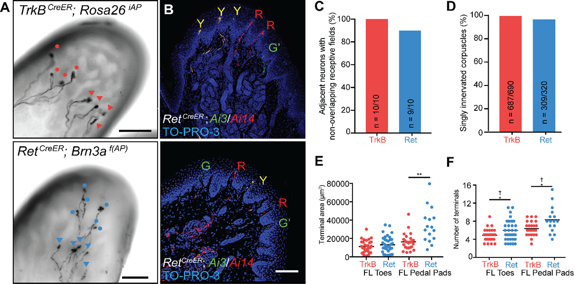
Fig. 5. Spatial arrangement of Ret+ and TrkB+ Meissner afferent cutaneous endings.

A. Images of whole-mount, AP-stained digital pads of a TrkBCreER; Rosa26iAP mouse (top) and a RetCreER; Brn3af(AP) mouse (bottom, Z-projection performed in Fiji) depicting pairs of afferents of the same subtype innervating the same glabrous pad. Tamoxifen doses were titrated to achieve sparse labeling of Meissner afferents. Terminals of one neuron are annotated with circles and terminals of the other with triangles. (scale bars = 100 μm) B. Digital pad sections of TrkBCreER; Rosa26LSL-YFP/LSL-tdTomato (Ai3/Ai14) mice treated with tamoxifen at E12.5 and E13.5 (upper panel) and RetCreER; Ai3/Ai14 mice administered tamoxifen at E11.5 and E12.5 (lower panel). Y: fibers express both tdTomato and YFP; R: fibers express tdTomato only; G: fibers express YFP only; G’: YFP+ fibers without terminals in the sections. Sections were stained with anti-DsRed, anti-GFP, and TO-PRO-3. (scale bar = 100 μm) C. Percentage of TrkB+ and Ret+ Meissner afferent pairs innervating the same pad and occupying different spatial territories. D. Quantification of the percentage of dermal papillae and Meissner corpuscles receiving a single TrkB+ fiber (3 animals) and a single Ret+ fiber (6 animals).
E. Receptive field surface area (terminal area, left) and number of terminal endings (right) of TrkB+ and Ret+ Meissner afferents measured in TrkBCreER; Rosa26iAP or TrkBCreER; Brn3af(AP) mice (N = 21) and RetCreER; Rosa26iAP or RetCreER; Brn3af(AP) mice (N = 21). Individual measurements and mean values (black bar) are plotted for forelimb digital pads and pedal pads. (two-tailed Welch’s t-test, mean significantly different: * p < .05, ** p < .01; F-test of the equality of variances, variance significantly different: † p < .01).
