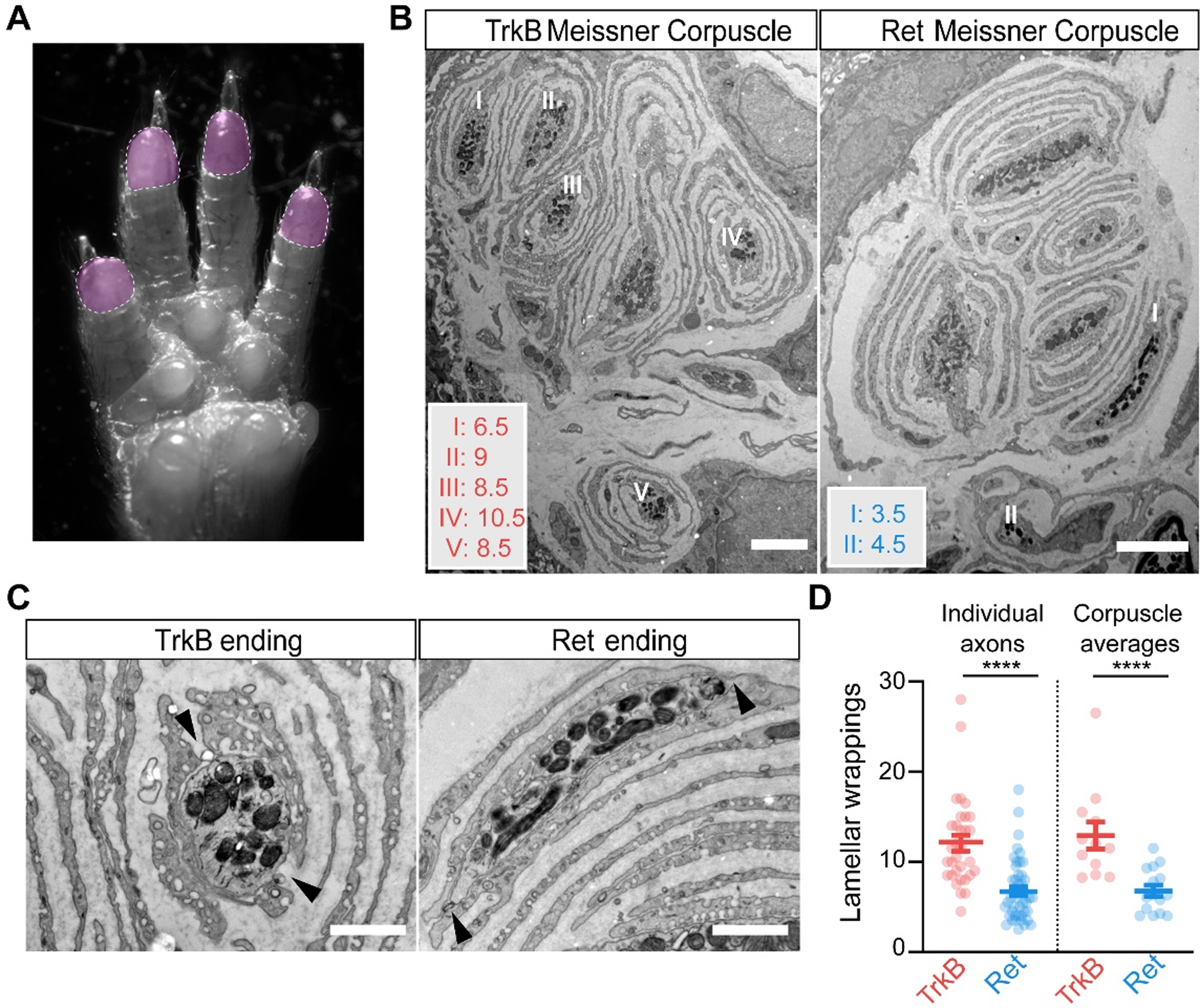
Fig. 6. TrkB+ Meissner Aβ LTMR endings have more lamellar cell wrappings than Ret+ Meissner mechanoreceptor endings.

A. Image of the paw, with the shaded areas (digit tips) representing regions of high density of Meissner corpuscles used for EM analysis. B. EM images of Meissner corpuscles from a TrkBCreER; AdvillinFlpO; Rosa26DR-Matrix-dAPEX2 mouse treated with tamoxifen at P3 (left) and a RetCreER; AdvillinFlpO; Rosa26DR-Matrix-dAPEX2 mouse treated with tamoxifen at E11.5 and P10 (right). Scale bar = 3 μm. C. High magnification images of labeled endings and associated lamellar cells from (B). Both exhibit openings or areas not directly associated with lamellar cells (black arrowheads). Scale bar = 1 μm. D. Quantification of the number of lamellar cell wrappings around genetically labeled endings of TrkB and Ret endings. Shown are the number of lamellar wrappings for individual axonal profiles (left panel; n = 32 and 47, respectively) and averages of all axonal profiles within individual corpuscles (right panel; n = 12 and 15, respectively). Mann-Whitney U test, **** p < 0.0001. N = 2 animals for each LTMR subtype.
