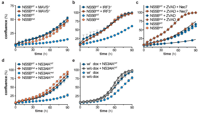
Figure 6.
Mitochondrial antiviral signaling protein (MAVS)- and interferon regulatory factor 3 (IRF3)-dependent negative selection of NS5B expressing cells and rescue by HCV NS3/4A. (a) Cell growth of wild-type and MAVS knock-out A549 cells, 5 days after transduction of either NS5BWT or NS5Bmut (mean ± s.d. of n = 5 technical replicates). One representative of n = 3 independent experiments is shown. (b) Cell growth of wild-type and IRF3 knock-out A549 cells, 5 days after transduction of either NS5BWT or NS5Bmut (mean ± s.d. of n = 5 technical replicates). One representative of n = 3 independent experiments is shown. (c) Cell growth of DMSO, Z-VAD-FMK (ZVAD, 40 µM), or Necrostatin-7 (Nec7, 10 µM) treated A549 cells, 5 days after transduction of either NS5BWT or NS5Bmut (mean ± s.d. of n = 4 technical replicates). One representative of n = 2 independent experiments is shown. (d) Cell growth of NS5BWT or NS5Bmut and NS3/4AWT or NS3/4Amut transduced A549 cells (mean ± s.d. of n = 3 technical replicates). One representative of n = 3 independent experiments is shown. (e) Cell growth of non-treated or dox-treated HEK-FlpIn-MAVS cells, 5 days after NS3/4AWT or NS3/4Amut transduction (mean ± s.d. of n = 9–12 technical replicates). One representative of n = 3 independent experiments is shown.

