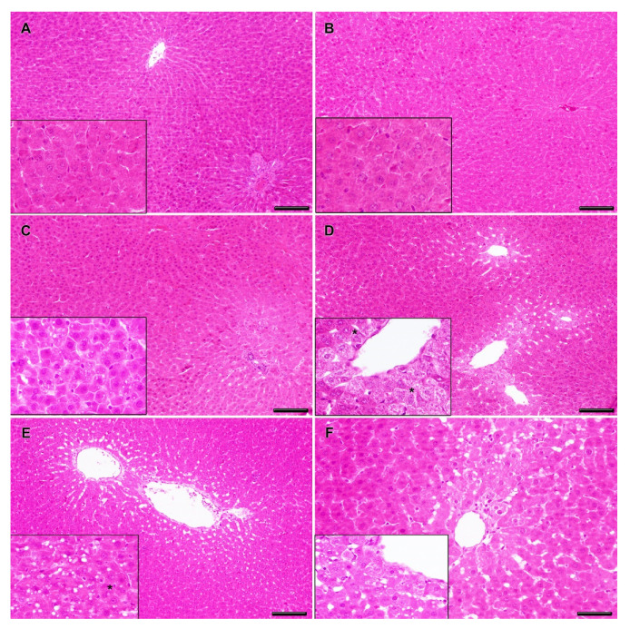
Figure 4.
Representative histopathological changes in the liver of rats exposed to CYN/MC-LR. Normal hepatic parenchyma is observed in negative (A) and solvent (B) control groups. Details of normal hepatocytes are observed in the insets (A,B). Rats exposed to 7.5 + 75 µg/kg b.w. CYN/MC-LR showed a diffuse distribution of mild degenerate hepatocytes with micro-vesicular lipid vacuolation (C). There are details of intracellular accumulation of small, round and clear vacuoles in hepatocytes (C, inset). Rats exposed to 23.7 + 237 µg/kg b.w. CYN/MC-LR presented mild degenerate and necrotic hepatocytes in centrilobular areas (D). There are details of degenerate hepatocytes, some of them multinucleated (*) (D, inset). Rats exposed to 75 + 750 µg/kg b.w. CYN/MC-LR showed moderate degenerate and necrotic hepatocytes in centrilobular areas with macro-vesicular lipid vacuolation (E). There are details of intracellular accumulation of large, round, and clear vacuoles inside hepatocytes, some of them multinucleated (*) (E, inset). The positive control group showed hepatocyte degeneration with lipid accumulation and centrilobular necrosis (F). There are details of the degeneration of hepatocytes with macro-vesicular lipid vacuolation (F, inset). Hematoxylin and eosin staining; bars = 100 μm (A–E) and 50 μm (F).

