Figure 1.
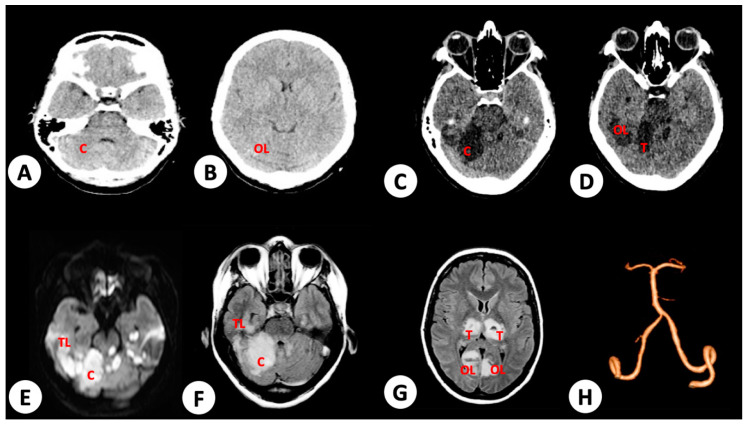
Brain radiologic imaging of patient 1. (A) No evidence of mass or vascular lesions in cerebellum or (B) occipital lobes (OL) in brain computed tomography (CT) immediately after seizure and decerebrate posturing events (10 days post onset of dengue symptoms). (C) T1 weighted image from brain nuclear magnetic resonance (NMR) performed 24 h after occurrence of disorientation and seizures, with evidence of ischemia in cerebellum (C) (11 days post onset of dengue symptoms). (D) Ischemic areas in thalamus (T) and occipital lobe. (E,F) Extensive ischemic infarct in cerebellum and parietal lobe (PL), in diffusion-weighted and fluid-attenuated inversion recovery (FLAIR) image from brain NMR performed 24 h after occurrence of acute clinical events (disorientation and seizures), respectively. (G) Ischemic infarct in thalamus and occipital lobe. (H) No evidence of vessel lesions, including basilar and vertebral arteries responsible to vascularization of injured areas in brain angiographic study performed 24 h after seizures.

