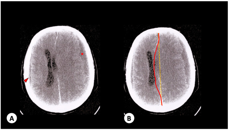
Figure 2.
Patient 2 radiologic observations. (A) Brain no-contrast computed tomography with hypodense images in right parieto-occipital (0.5 cm—arrowhead) and left front-temporal (1.6 cm—red star) areas. (B) Corresponding brain contrasted computed tomography with middle-line deviation (red line) compared to regular condition (yellow line). It is not possible to view left lateral ventricular and cerebellar cisterns due to compression by left subarachnoid hemorrhage.

