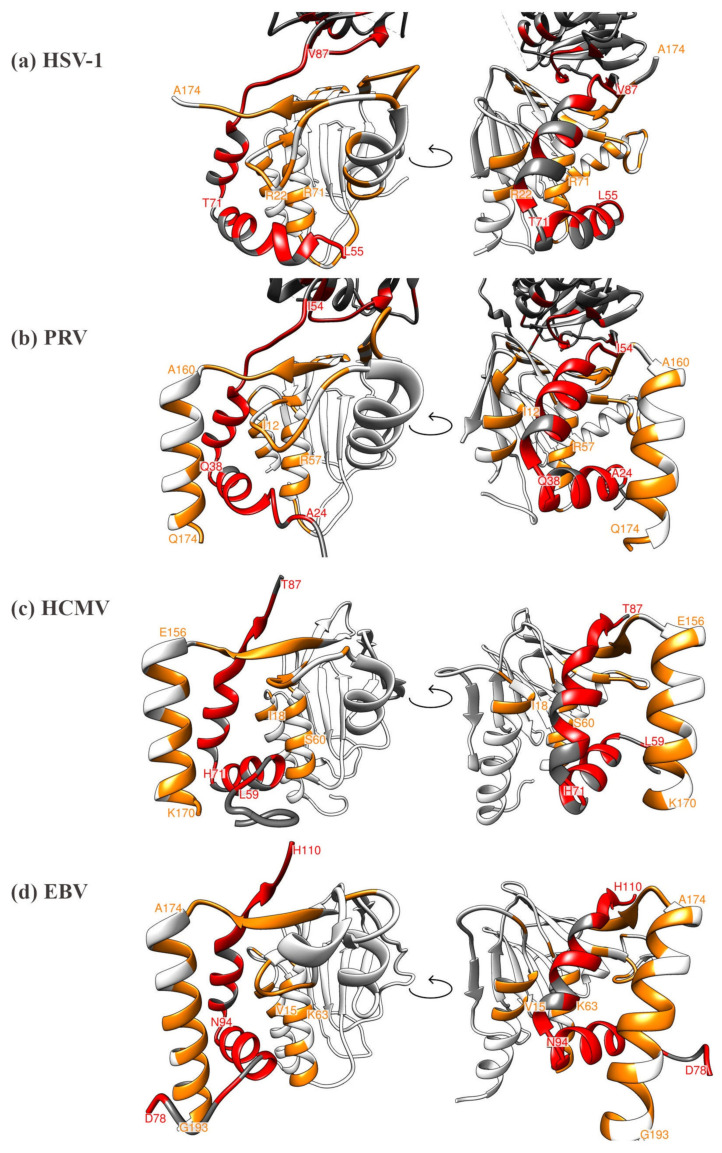
Figure 4.
Comparison of the currently available four herpesviral 3D core NEC crystal structures, highlighting the heterodimeric contact interfaces, each in two different viewing angles (left and right rotated by 90 degrees). (a) HSV-1 core NEC, (b) PRV core NEC, (c) HCMV core NEC, (d) EBV core NEC. Interacting residues within the hook or groove are colored in red and orange, respectively. Distinct amino acids marking the start or end positions of structural elements are labeled as orientation points.

