Figure 5.
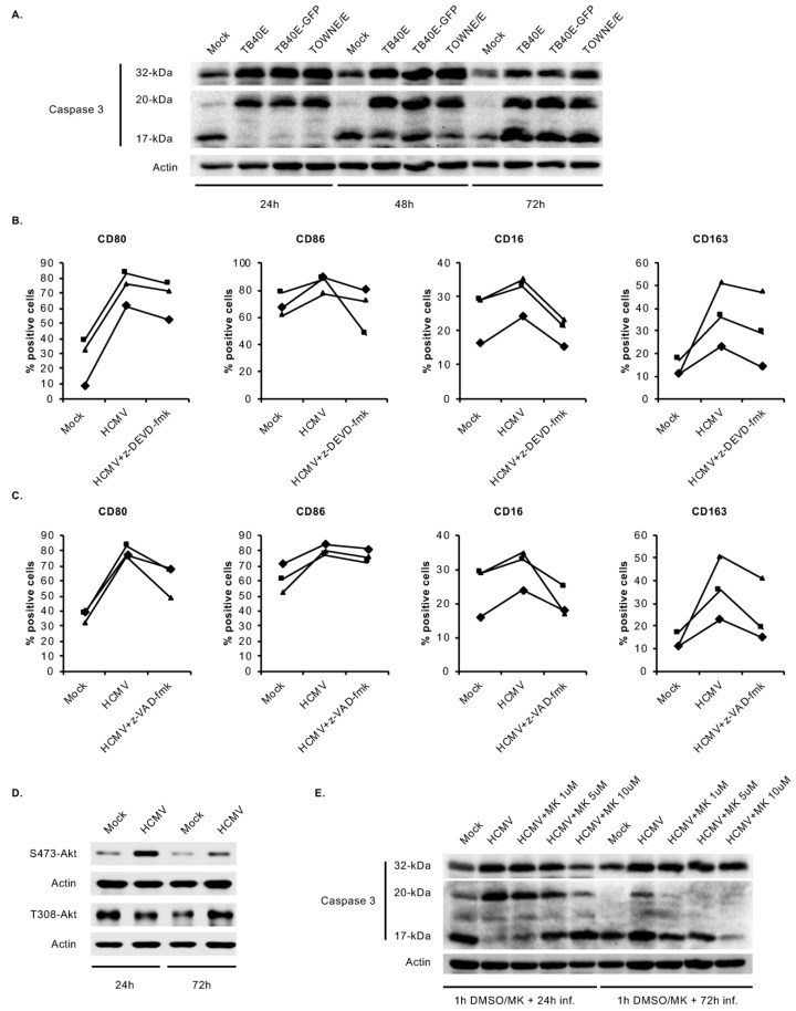
HCMV-induced caspase 3 activation after the 48-h viability gate mediates the unique monocyte-to-macrophage differentiation. (A) Human peripheral blood monocytes were mock- or HCMV-infected with TB40E, TB40E-GFP or TOWNE/E for 24, 48, or 72 h. Levels of pro-caspase 3 (32 kDa), intermediate caspase 3 (20 kDa), fully active caspase 3 (17 kDa), and actin were detected by immunoblotting from whole cell lysates. (B,C) Monocytes were pretreated for 1 h with a vehicle control, (B) a caspase 3 inhibitor (z-DEVD-fmk) at 20 μM, or (C) a pan-caspase inhibitor (z-VAD-fmk) at 50 μM. Cells were then mock- or HCMV-infected for 48 h and the percent positive cells for M1 (CD80 and CD86) and M2 macrophage markers (CD16 and CD163) were measured by flow cytometry. (D) Monocytes were mock- or HCMV-infected for 24 or 72 h. Levels of S473-Akt, T308-Akt, and actin were detected by immunoblotting. (E) Monocytes were pretreated for 1 h with MK at 1, 5, or 10 μM, or vehicle control and then mock- or HCMV-infected for 24 h or 72 h. Levels of pro-caspase 3 (32 kDa), intermediate caspase 3 (20 kDa), fully active caspase 3 (17 kDa), and actin were detected by immunoblotting. (A–E) Results are representative of 3–6 independent experiments using monocytes from different donors.

