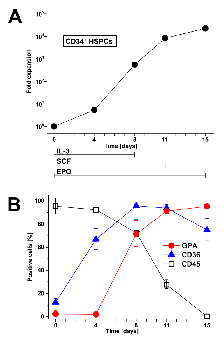
Figure 3.
Cytokine-driven amplification of ex vivo cultured CD34+ HSPCs monitored in a time interval of 15 days (A) and the portrayal of cell surface markers of the developing erythrocytes (B). Cells were cultivated in the presence of interleukin (IL)-3, stem cell factor (SCF), and erythropoietin (EPO), which were applied from the initiation of cultivation at day 0 until day 8 (IL-3), day 11 (SCF), and the end of cultivation at day 15 (EPO). The graph shows the average cell expansion of HSPCs of three donors ([145], modified).

