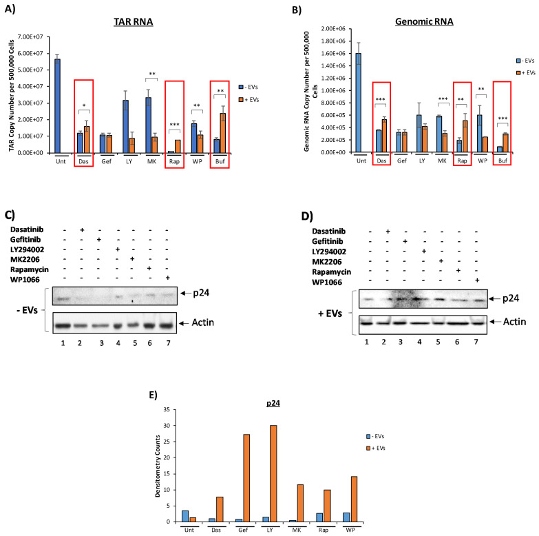
Figure 3.
Effect of EVs on the activation of HIV-1 in the presence of various kinase inhibitors. CEM EVs were isolated by ultracentrifugation. Here, 5 × 10⁵ U1 cells were plated and treated with 5 µM dasatinib, 10 µM gefitinib, 10 µM LY294002, 1 µM MK2206, 150 nM rapamycin, or 1 µM WP1066 and allowed to incubate for 48 h. A second drug treatment was then performed. This was followed by a 2-h incubation and a CEM EV treatment. A second EV treatment was performed after a 24-h incubation period. The total ratio of cells to EVs was 1:10,000. Cells were then allowed to incubate for 24 h prior to harvest. Cell supernatant was collected and rotated overnight at 4 °C with NT86. Total RNA was isolated and subjected to RT-qPCR for HIV-1 TAR RNA (A) and genomic RNA (B). Red boxes indicate increased HIV-1 TAR and genomic RNA levels in EV-treated cells in the presence of dasatinib, rapamycin, and bufalin. (C,D) NT86-treated samples were Western blotted for HIV-1 Gag p24. U1 WCE was used as a positive control. Actin was used as a loading control. (E) Densitometry counts normalized to actin are shown for HIV-1 Gag p24. For all figures, EV untreated samples were used as negative controls. Student’s t-test compared untreated cells with cells treated with drugs. *, p < 0.05; **, p < 0.01; ***, p < 0.001. Error bars, S.D.

