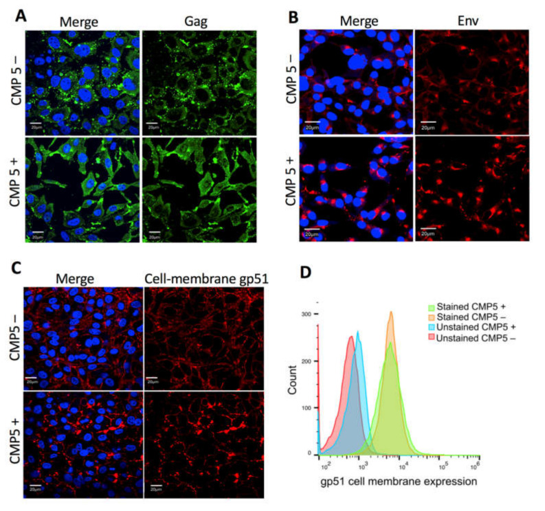
Figure 8.
Effect of CMP5 on Gag and Env localization. (A,B) FLK-BLV were grown on coverslips and treated with Milli-Q water (CMP5–) or CMP5 20 µM (CMP5+), Gag was stained with green fluorescence (A), Env was stained with red fluorescence (B), The cellular membrane staining of gp51 was performed without the permeabilization step (C). DAPI (blue fluorescence) was used to stain the nucleus and the merge picture represents Gag or Env with DAPI. The data are a representation of three experiments. Images were acquired with a 60X objective and the scale bar is equal to 20 µm. (D) FLK-BLV were treated with Milli-Q water (CMP5–) or CMP5 20 µM (CMP5+), the cells were collected, and gp51 cell membrane expression was assessed by flow cytometry. The data are a representation of two experiments. Scale bar—20 µm.

