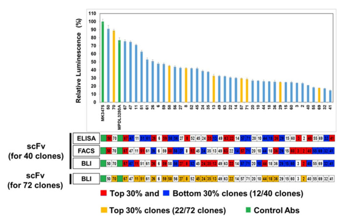
Figure 4.
In vitro cell-based PD-1/PD-L1 inhibition assay in comparison with primary scFv screenings. Evaluation of in vitro assays to assess the inhibition of PD-1‒PD-L1 interactions by full IgG antibodies. The activity of 40 full IgG converted antibodies in blocking the effect of the PD1/PD-L1 checkpoint on TCR-mediated T cell activation is assessed as the level of luciferase activity. MK3475 (anti-PD-1) and MPDL3280A (anti-PD-L1) antibodies were used as positive controls (green bar). The light blue bars have binding activity of antibody clones against human PD-L1 antigen, whereas yellow bars have cross-reactivity to both human and mouse PD-L1 antigens. Results are classified using color shading codes, with the top 30% in red and the bottom 30% in blue in each assay (scFvs 30%; 12/40 clones and 22/72 clones).

