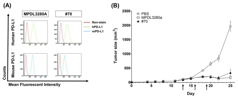
Figure 6.
In vivo function of anti-PD-L1 antibody in MC38 syngeneic mouse model. (A) Flow cytometry analysis demonstrates cross-reactive binding of IgG-converted anti-human PD-L1 antibodies to human PD-L1 in PC3 cells, and mouse PD-L1 in mouse IFN-γ-treated MC38 tumor cells (100 ng/mL for 24 h); (B) Tumor growth curves of C57BL/6 mice subcutaneously injected with MC38 tumor cells. Mice (C57BL/6 bearing MC38 tumors, n = 5 per group, mean ± SD) were treated with PBS or mouse PD-L1 cross-reactive antibodies (MPDL3280A and #70). Treatment was given by intraperitoneal injection (10 mg/kg) on the days marked with black arrows. t-test; ***; p ≤ 0.001 by unpaired t-test on day 25.

