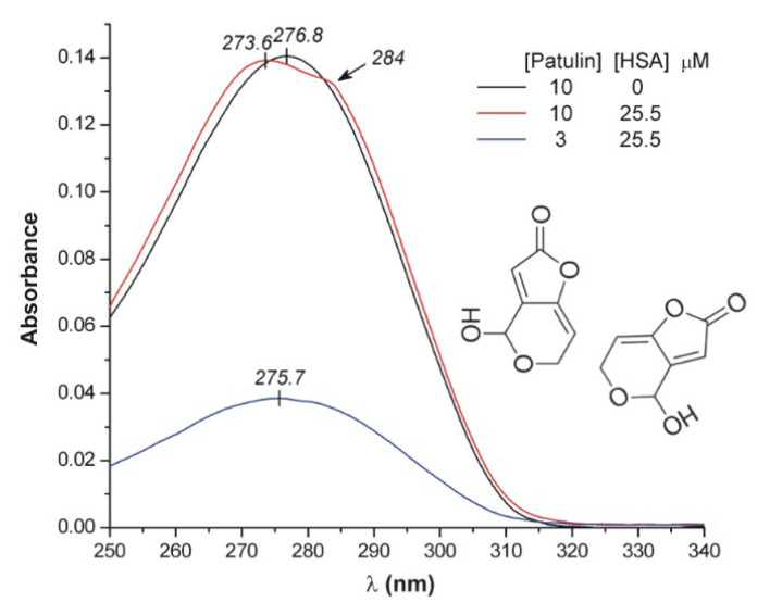
Figure 7.
Comparison of the UV absorption spectra of patulin (PAT) recorded in the absence and presence of 25 μM human serum albumin (HSA; pH 7.4, 25 °C, optical path length: 1 cm). A possible relative steric arrangement of two PAT molecules at the HSA binding site is shown. Long axes of the conjugated π-systems are nearly perpendicular to each other, resulting in the exciton splitting of the UV band.

