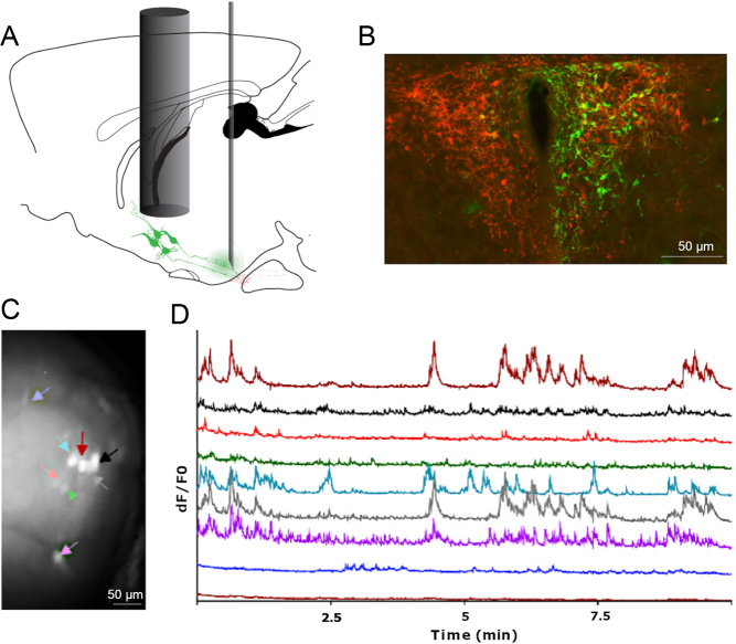
Figure 3.
In vivo calcium imaging of TRH neurons in conscious mice. (A) Trh-IRES-Cre mice were injected in the median eminence with a viral vector (AAV5.CAG.Flex.GCaMP6s.WPRE.SV40) and TRH neurons were visualised through a GRIN lens placed above the PVN. (B) Immunofluorescence image showing GCaMP6s expression (green) in TRH neurons (red) located in the PVN. (C) GRIN lens view of a field of the TRH neurons expressing GCAMP6m. Regions of interest corresponding to individual neurons are indicated by coloured arrows. (D) Time-lapse recordings of calcium (GCAMP6s) activity of eight hypophysiotropic TRH neurons in an adult Trh-IRES-Cre male mouse. Scale bars 50 μm.

 This work is licensed under a