Figure 2.
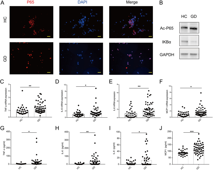
NF-κΒ signaling pathway is activated in GD patients. (A) Immunofluorescence of PBMCs with P65 (red) and DAPI (blue) (n = 5). Scale bars, 20 μm. (B) Western blotting analysis of key molecules of the NF-κB pathway in GD patient and HC PBMCs. (C–F) The mRNA expression of TNF-α (C), IL-6 (D), IL-8 (E) and MCP1 (F) was measured by qRT-PCR in GD patient (n = 51) and HC (n = 30) PBMCs. (G–J) The serum levels of TNF-α (G), IL-6 (H), IL-8 (I) and MCP1 (J) were measured by ELISA in the patients with GD (n = 51) and HCs (n = 30). GD, Graves’ disease; HC, healthy control. Data represent means ± s.e.m. *P < 0.05, **P < 0.01, ***P < 0.001.

 This work is licensed under a