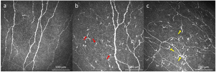
Figure 5.
In vivo confocal microscopy images in dry eye disease: a) Healthy individual. b) Increased number of dendritiform cells in patients with dry eye disease (red arrows show representative cells). c) Increased number of presumed mature dendritiform cells in patient with dry eye disease (yellow arrows show representative cells).

