
. 2020 Mar 12;7(2):G51–G58. doi: 10.1530/ERP-19-0060
 This work is licensed under a
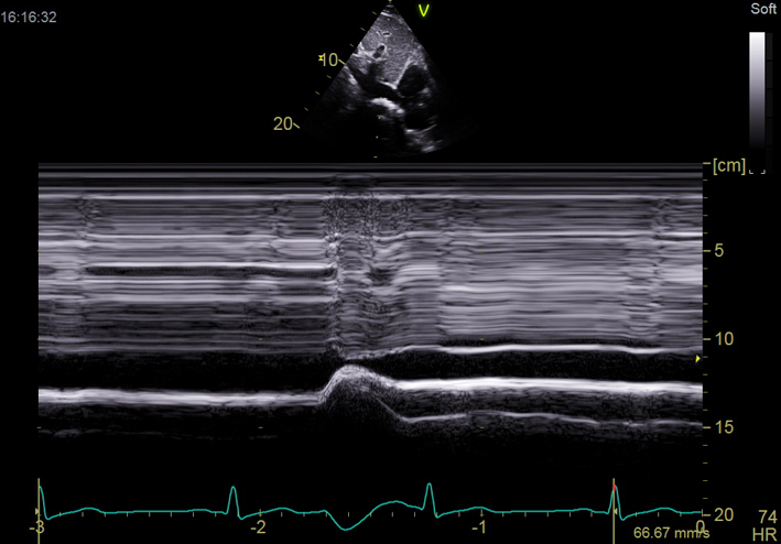
 Table 1.
The minimum data set for a Level 1 echocardiogram.
| View/modality | Explanatory note | Image |
|---|---|---|
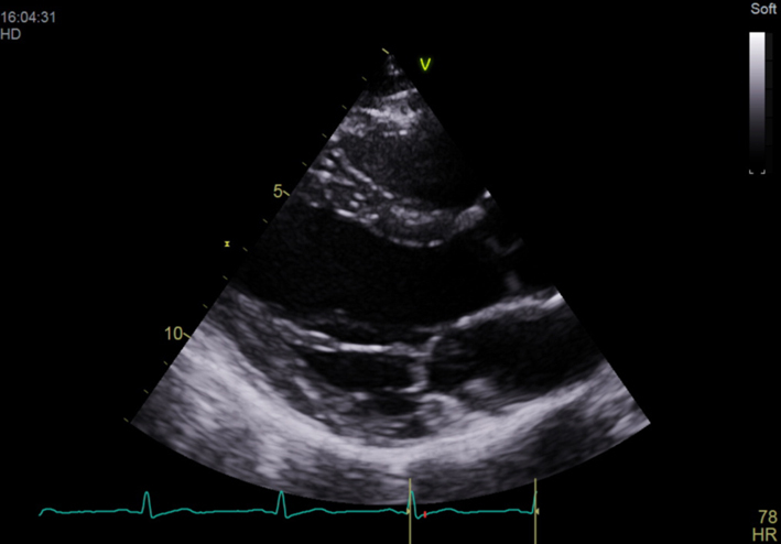
| PLAX deep (2D) | Visual assessment of pleural and pericardial effusions | 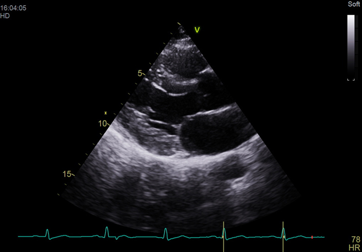 |
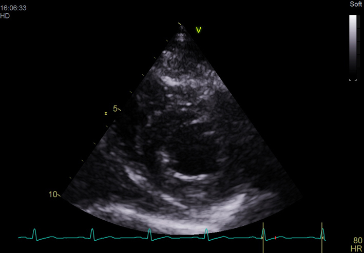
| PLAX (2D) | Optimise depth to look at cardiac structures Visual assessment of LV size and function Visual assessment of RV AV appearance and movement MV appearance and movement Major regional wall motion abnormalities |
 |
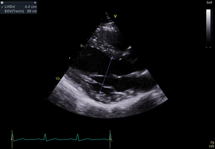
| PLAX (2D) LVIDd | Measurement of left ventricular internal diameter (LVID) in diastole Visual assessment of aortic root size |
 |
| PLAX (2D)with colour Doppler over aortic valve (AV) | Check colour Doppler settings Colour box over AV Look for abnormal flow over AV |
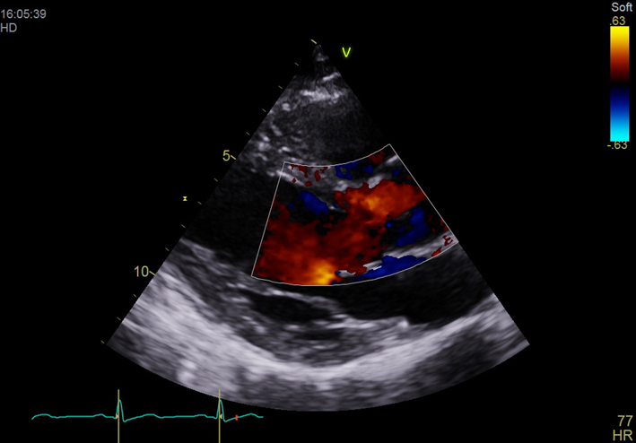 |
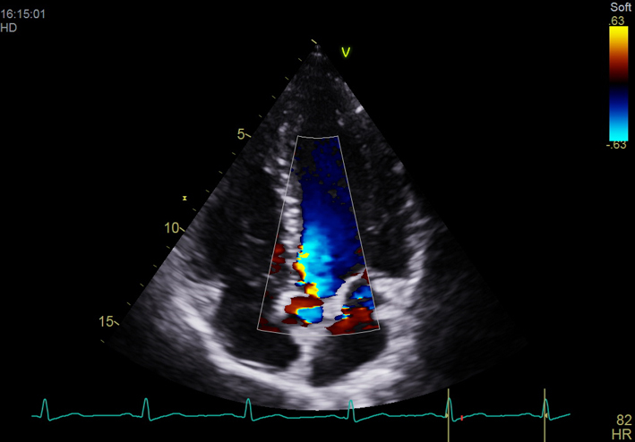
| PLAX (2D) with colour Doppler over mitral valve (MV) | Check colour Doppler settings Colour box over MV Look for abnormal flow over MV |
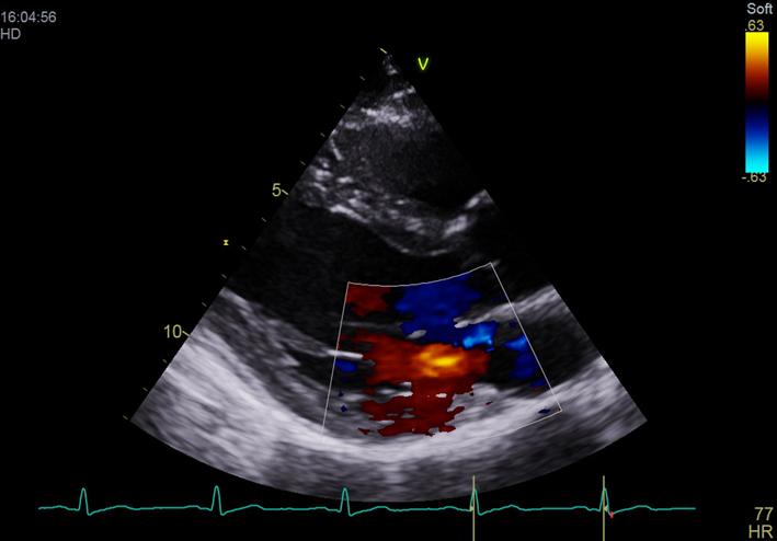 |
| PSAX Outflow (2D) | AV appearance and function | 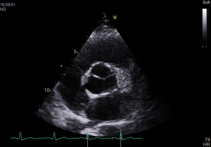 |
| PSAX Base (2D) | MV appearance and function Radial systolic function Major regional wall motion abnormalities |
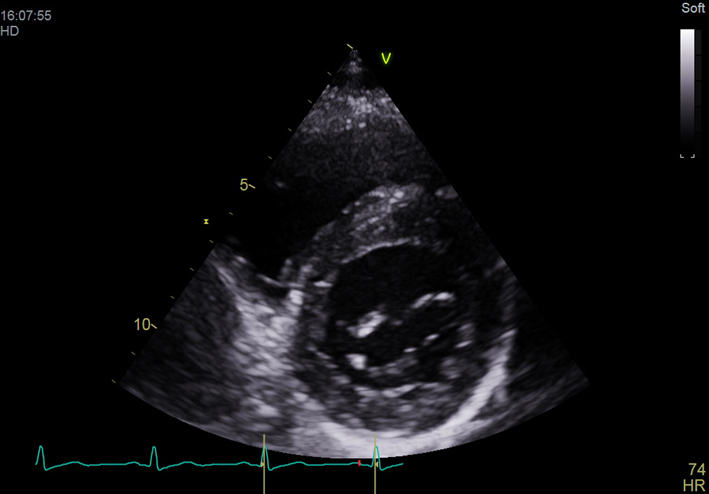 |
| PSAX Mid (2D) | Radial systolic function Major regional wall abnormalities |
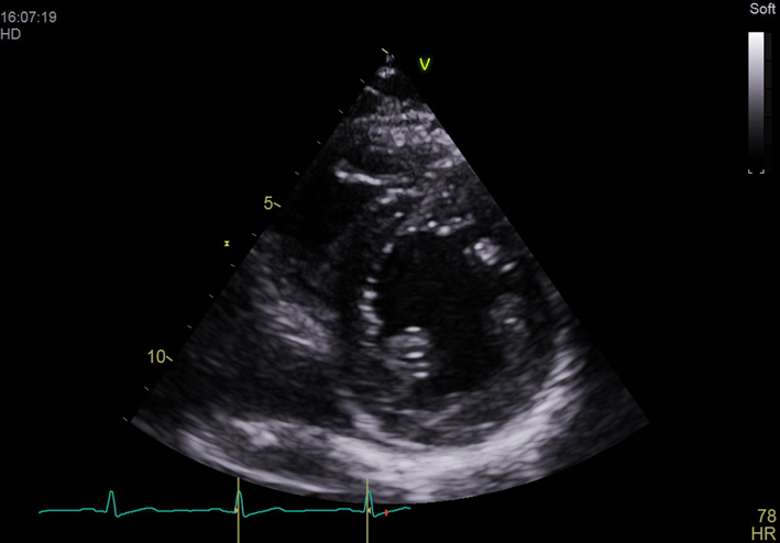 |
| PSAX Apex (2D) | Radial systolic function Major regional wall abnormalities |
 |
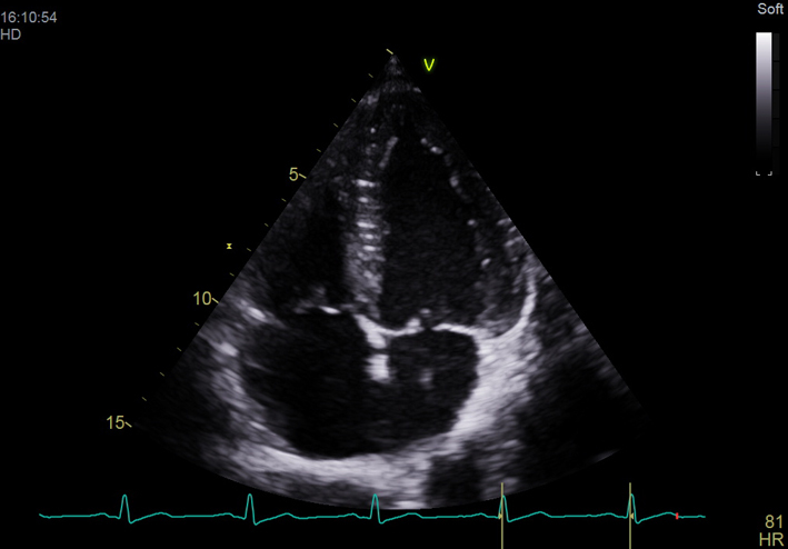
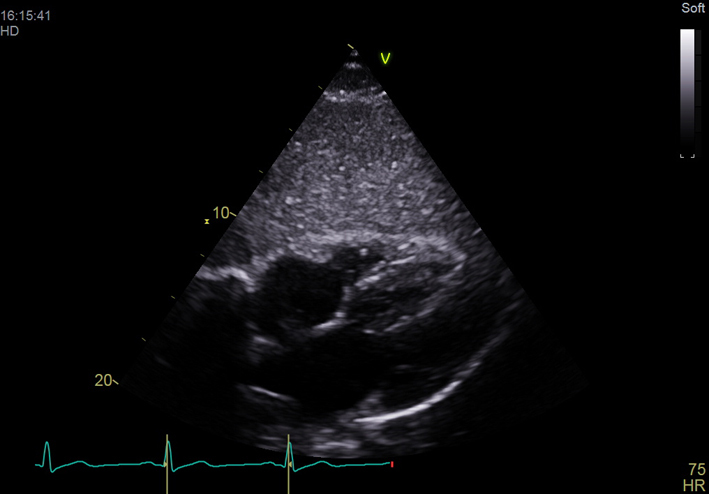
| Apical 4 chamber (2D) | LV cavity size (visual assessment) Visual assessment of longitudinal and radial function Major regional wall abnormalities MV appearance and function TV appearance and function RV cavity size (compared with LV size) RV free wall motion (visual assessment) Visual assessment of RV free wall hypertrophy Observe atrial septal position/mobility |
 |
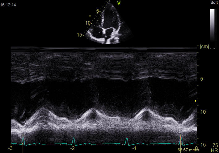
| A4C (M-Mode) TAPSE | M-mode through the lateral tricuspid valve annulus Measure tricuspid annular plane systolic excursion (TAPSE) |
 |
| A4C (2D) with CFM TV | Check colour Doppler settings Look for abnormal colour flow over TV |
 |
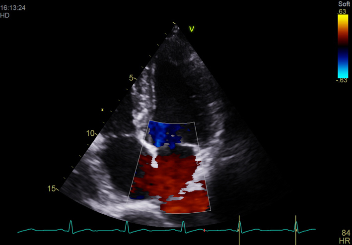
| A4C (2D) with CFM MV | Check colour Doppler settings Look for abnormal colour flow over MV |
 |
| A5C (2D) | AV appearance and function | 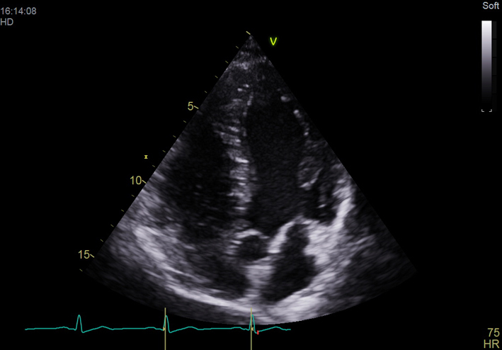 |
| A5C (2D) with CFM AV | Check colour Doppler settings Look for abnormal flow over AV |
 |
| Subcostal 4Ch (2D) | Relative chamber sizes and function Visual assessment of RV free wall hypertrophy Atrial septal position and hypertrophy Pericardial effusion |
 |
| Subcostal IVC (2D) | Observe size and collapsibility with respiration | 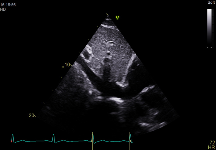 |
| Subcostal IVC (M-mode) | Assess IVC size and variation with respiration Dilated IVC may indicate high right atrial pressure |
 |