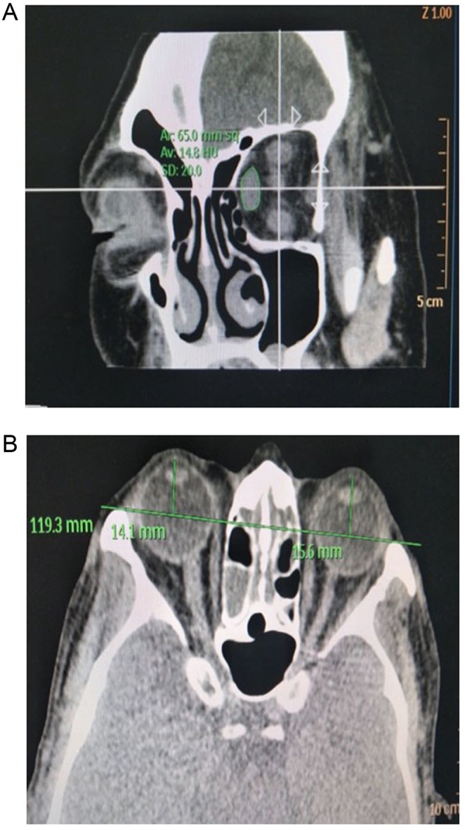
Figure 1.

Coronal and axial CT images of a representative patient with GO. (A) Measurement of the cross-sectional area of the OM and the orbit. On the coronal plane of CT scan of the orbit, with the back of the eyeball as the tangent line, layer thickness 1 mm, and pitch 1 mm, 2-mm plane was found behind the ball. Superior, lateral, inferior, and medial extraocular muscle areas were observed. The cross-sectional area of the orbit and each extraocular muscle was measured by AutoCAD software, and each extraocular muscle was measured three times to obtain an average value. (B) Measurement of exophthalmometry. Exophthalmometry was measured according to the standard that the center of the lens and the intraorbital segment of the optic nerve are displayed on the same side and the outer edge of the orbit was at the lowest point.

 This work is licensed under a